Technological advancements have enabled the dental field to make remarkable progress, enhancing patient care and diagnostic precision. One of the most transformative innovations is digital radiography, which has replaced traditional X-ray film as the standard in modern dental imaging.
Traditional X-rays were once a source of concern for patients because of radiation exposure and the discomfort of biting on sharp film packets. Today, digital X-rays have changed everything. They provide clear images, use significantly less radiation, and allow your dentist to detect oral health issues earlier than ever before.
At Dr. Fred Alger Periodontics & Dental Implants, we rely on advanced digital imaging systems to monitor and diagnose your oral health safely and effectively. Dr. Alger believes in using state-of-the-art dental technology whenever possible to improve treatment planning, accuracy, and patient comfort.
What Are Digital X-rays?
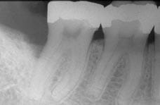
Post-Treatment
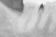
Pre-Treatment
Seeing the bone and root structures under the gums is vital for accurate diagnoses. Digital X-rays use electronic sensors instead of traditional film to capture detailed images of your teeth, jaw, and surrounding structures. These images are processed instantly in a digital format, eliminating the need for chemical processing or physical film.
Dr. Alger utilizes this technology to detect gum disease, bone loss, tooth decay, and subtle changes in ligament or bone density that may indicate infection or other underlying medical conditions. Because the images appear instantly on a computer screen, he can zoom in on specific areas, improving precision and helping you visualize exactly what’s happening in your mouth.
Since their development, digital X-rays have proven far superior to traditional photographic film, offering greater precision, quicker results, and enhanced patient safety. They reduce radiation exposure by up to 90% compared to older systems, making them a safer option for routine and advanced dental care.
How Digital Radiography Works
During your visit, one of our dental assistants will place a small digital sensor or flat panel detector in your mouth to capture the image. The process takes only seconds, and the data is instantly transmitted to a computer monitor in the treatment room.
Because the system uses electronic sensors, there’s no waiting for film development or chemical processing. This means you and Dr. Alger can review your X-rays immediately, allowing for a faster diagnosis and a more efficient treatment plan.
You’ll also wear a lead apron for additional protection, even though modern systems already use less radiation than most household electronics. This commitment to safety ensures peace of mind while maintaining diagnostic excellence.
Key Advantages of Digital Radiography
The shift from traditional film to digital radiography has revolutionized dental care in numerous ways. Some of the key advantages include:
- Reduced radiation exposure: Digital systems require up to 90% less radiation than conventional X-rays.
- Immediate availability of images: Results appear on-screen within seconds for real-time review.
- Enhanced image quality: Digital images provide incredible detail, allowing dentists to identify subtle changes that would go unnoticed on older films.
- Better treatment planning: Images can be magnified, color-adjusted, or enhanced for more precise evaluation.
- Eco-friendly process: Digital systems eliminate the chemicals and waste produced by film processing.
- Improved patient communication: Dr. Alger can show you your own images of your teeth and explain areas of concern directly on-screen.
- Continuity of care: Files can be securely shared with your general dentist or other healthcare providers, ensuring collaborative, consistent treatment.
Because of these advantages, digital imaging has become the standard in modern dental practices and a vital component of our commitment to delivering the highest level of care.
Digital X-rays and Oral Health Monitoring
Regular digital radiographs are crucial for the early detection of dental issues, particularly for patients with a history of periodontal disease or decay. These detailed images help identify bone loss, infections, or tooth fractures before they cause pain or require more complex dental procedures.
Dr. Alger recommends incorporating digital X-rays as part of your regular dental exam to maintain optimal oral health. They help detect:
- Early gum disease and bone deterioration
- Infections beneath the gum line
- Impacted or misaligned teeth
- Complications related to dental implants or restorations
- Changes in bone density over time
This ability to identify problems at their earliest stages supports effective care, faster recovery, and improved patient outcomes.
How Digital Imaging Enhances Treatment Planning
Whether preparing for implant placement, orthodontic planning, or periodontal surgery, digital X-rays provide the detailed insights Dr. Alger needs for precise and predictable results. The clarity of digital images enables more accurate measurements, ensuring the precise placement of dental implants and protecting vital structures.
Because images can be saved and compared over time, digital radiography also enables Dr. Alger to track healing progress, monitor bone growth, and evaluate the success of past procedures. This continuity is key to long-term success and improved quality of life for every patient.
The Role of Digital Radiography in Patient Education
One of the most valuable aspects of digital imaging is how it improves patient understanding. Seeing images of your mouth in real time helps you visualize your condition and feel more involved in your treatment options. Patients often find it easier to understand the need for certain procedures, such as deep cleanings, bone grafts, or periodontal therapy, when they can see the issue firsthand.
Dr. Alger and his team use these visuals as a teaching tool, helping you make informed decisions about your oral health and ongoing dental care. It’s part of our promise to provide transparent, personalized, and compassionate care for every patient.
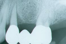
The bicuspid tooth supporting this upper left bridge had lost 50% of the bone around the root. Harmful biting forces may have been a contributing factor to the bone loss observed. A removable partial denture or extraction and replacement with implants were options available. However, this lady wanted to keep her own teeth, if at all possible.
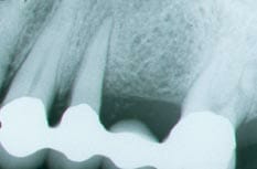
Dr. Alger was able to regenerate bone around the tooth with a bone grafting procedure. Afterwards her dentist replaced her old bridge with a new one that provided better chewing stability.